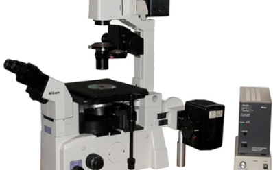
We use confocal microscopy to image the distribution of fluorescent molecules in the vesicle membrane.
The microscope chassis is the inverted TE2000 Nikon. It is equipped with both traditional bright field, and epifluores- cence modules. The following objectives are available: x10, x20, x40 DIC, x40 Phase Contrast, x60 WI DIC, x100 OIL. The confocal module is available with the following laser wavelengths: 408 nm, 456-477-488-514 nm, and 543 nm. Images at three wavelengths can be acquired simultaneously, using three PMTs. Alternatively, images can be acquired in the ’spectral mode’, using a unique 32-channel PMT spectral detector, bandwidth per channel: 2.5, 5, or 10 nm.
Home > RESEARCH > Systems & Techniques > Confocal Microscopy

